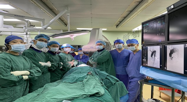
Recently, the minimally invasive aortic team of the Department of Cardiovascular Surgery of West China Hospital, Sichuan University, performed “total endovascular aortic repair using physician-modified endograft with triple inner branches for extensive arch aneurysm after ascending aorta replacement” for a 69-year-old male. The patient was discharged without complications on the 4th day after the operation. The literature review showed that no similar surgical case has been reported in China.
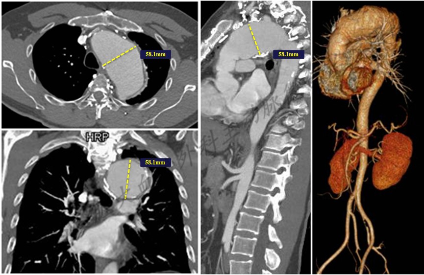
The patient, Mr. Wu, with persistent chest pain and progressively worsen hoarseness was admitted to WCH. Past medical history was notable for previous ascending aorta replacement for Debakey II dissection 5 years ago. CT angiography demonstrated an extensive arch aneurysm with a maximum diameter of 57.1 mm in short-axis (Fig. 1). The aortic arch lesions need to be solved urgently; otherwise, it would be progressed to devastating aortic dissection or even aortic rupture. However, the huge trauma and surgical risks from rethoracotomy daunted the patient and his families, and they even considered giving up the surgical treatment.

However, general endovascular strategies conventional branch reconstruction? such as parallel stents, in-vitro pre-fenestration and in-situ fenestration are also exposed to high risks of endograft-related complications. For example, such effort won’t make sense for the patient once endoleaks occur. After aortic team discussion, the patient was accepted for a total endovascular approach by using physician modified endograft with triple inner-branch to reduce potential risk of resternotomy, peri-procedural cerebral ischemia and endograft-related complications, which succeeded eventually.
Aortic arch disease is a kind of cardiovascular disease that seriously affects the long-term prognosis of patients. Traditional median thoracotomy, especially rethoracotomy, makes patients exposed to high risks. Total endovascular repair can isolate the lesion site as minimally-invasive as possible. However, an individualized and accurate plan should be formulated based on the aortic anatomy and lesion characteristics of patients.